
Figure 7.
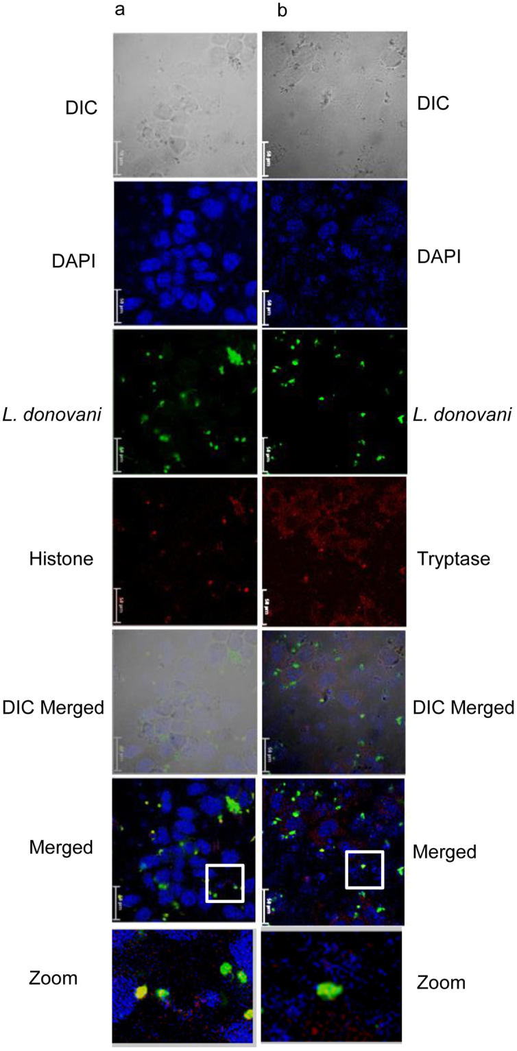
Immunostaining of MCETs with histone and tryptase on in vitro interaction of RBL MCs with L. donovani. Cells cultured on a cover glass slip for overnight incubated with L. donovani (green) for 24 h at an MOI of 1:10 in RBL medium and were processed as discussed in materials and methods. Panel a represent staining with anti – histone and panel b represent staining with anti – tryptase and were visualized under confocal microscopre at 60X. Scale bar is 50 μm, n = 3.