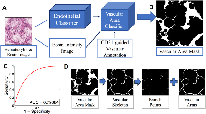
Figure 1.
Vascular area delineation in H&E stained slides using sequential, machine learning-based, 2-step vascular area classification approach. (A) Training of the first classification model: The original H&E image is processed into an endothelial nuclear mask based on hematoxylin staining and an eosin intensity image. The digital image of CD31 immunohistochemistry provides vascular annotation to train the vascular area classifier. (B) The second classifier outputs a vascular area mask (VAM) where vascular areas are white, and non-vascular areas are black. (C) Receiver Operating Characteristic (ROC) curve for vascular area classification compared to the vascular annotation provided by CD31 immunohistochemistry (AUC = 0.78). (D) Post-processing of the VAM.

