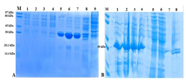
Fig. 3.

SDS-PAGE of HspX/EsxS fusion protein before and after renaturation with 3 and 6 M guanidine HCl. Gels were stained with Coomassie Brilliant Blue. A) HspX/EsxS fusion protein expression in E. coli strain BL21. Lane M: protein size marker; lanes 1 to 4: supernatant of BL21 lysate in different concentration of IPTG (0.1, 0.5, 1 and 2 mM); lanes 5 to 8: sediment of BL21 lysate in different concentration of IPTG (0.1, 0.5, 1 and 2 mM); lane 9: E. coli strain BL21 without HspX/EsxS fusion protein. B) Refolded fusion protein using 3 and 6 M guanidine HCl. Lane M: protein size marker; lanes 1 and 2: supernatant of BL21 lysate treated with 6 M guanidine HCl; lanes 3 and 4: supernatant of BL21 lysate treated with 3 M guanidine HCl; lanes 5 and 6: pellet of BL21 lysate treated with 6 M guanidine HCl; lanes 7 and 8: pellet of BL21 lysate treated with 3 M guanidine HCl.
