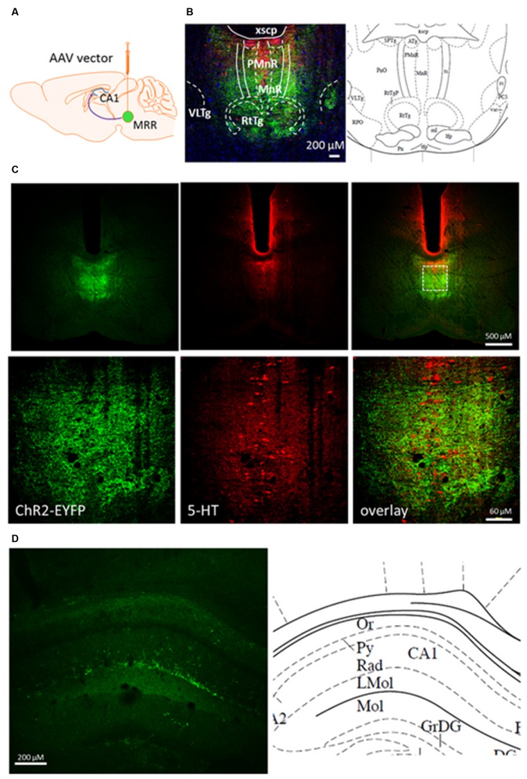
FIGURE 1.
Coronal section from pontine area and dorsal hippocampus of rAAV-injected mouse brain 8 weeks after. (A) Mice were injected with viral vector in the MRR. (B) Immunohistochemically enhanced EYFP marker protein (green) and 5-HT containing cells (red) in the raphe region (–4.60 mm from Bregma) by confocal microscopy. Cell nuclei (blue) was stained by Hoechst 33258. (C) In an injected mouse, 5-HT (red) is expressed in many EYFP-labeled neurons (green) along the midline. Under, show the zoom-in view of the dashed rectangular area. (D) EYFP containing neuronal projections (green) originate from the MR in the str. radiatum of dorsal part of CA1, –1.82 mm from Bregma. Corresponding regions and abbreviations of typical nuclei in the given area shown from The Mouse Brain by Paxinos (Paxinos and Franklin, 2012). Lmol, lacunosum moleculare layer of the hippocampus; MnR, median raphe nucleus; Or, oriens layer of the hippocampus; PMnR, paramedian raphe nucleus; Py, pyramidal cell layer of the hippocampus; Rad, stratum radiatum of the hippocampus; RtTg, reticulotegmental nucleus of the pons; VLTg, ventrolateral tegmental area; xscp, decussation of the superior cerebellar peduncle. (A) was created by modifying images purchased in the PPT Drawing Toolkits-BIOLOGY Bundle from Motifolio, Inc. (http://www.motifolio.com/neuroscience.html).

