Figure 1.
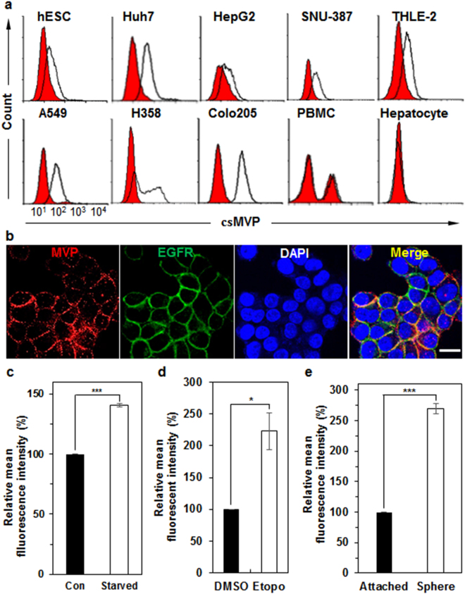
csMVP is expressed on HCC cells but not on normal hepatocytes, and is induced under stressful environments, such as serum starvation, DNA damage, and detachment stress. (a) Flow cytometry analysis of HCC cells (Huh7, HepG2, and SNU-387), immortalized hepatocytes (THLE-2), NSCLC cells (A549 and H358), colorectal cancer cells (Colo205), PBMCs, and primary hepatocyte with α-MVP. (b) Immunocytochemical analysis of Huh7 cells with α-MVP and EGFR antibodies. Huh7 cells were incubated with α-MVP (red) and anti-EGFR (green) antibodies. Nuclei were labelled with DAPI. The scale bar is 20 μm. (c) Induced expression of csMVP on the surface of serum-starved Huh7 cells. Relative expression of csMVP was measured by mean fluorescence intensities (MFIs) of flow cytometric analysis of normal (con) or serum-starved Huh7 cells (starved). ***p < 0.005. (d) Induced expression of csMVP on the surface of etoposide-treated Huh7 cells. Relative expression of csMVP was measured by MFIs of flow cytometric analysis of DMSO or Etopo-treated Huh7 cells. *p < 0.05. (e) Induced expression of csMVP on Huh7 sphere cells. Relative expression of csMVP was measured by MFIs of flow cytometric analysis of attached or sphere cultures of Huh7 cells. ***p < 0.005.

