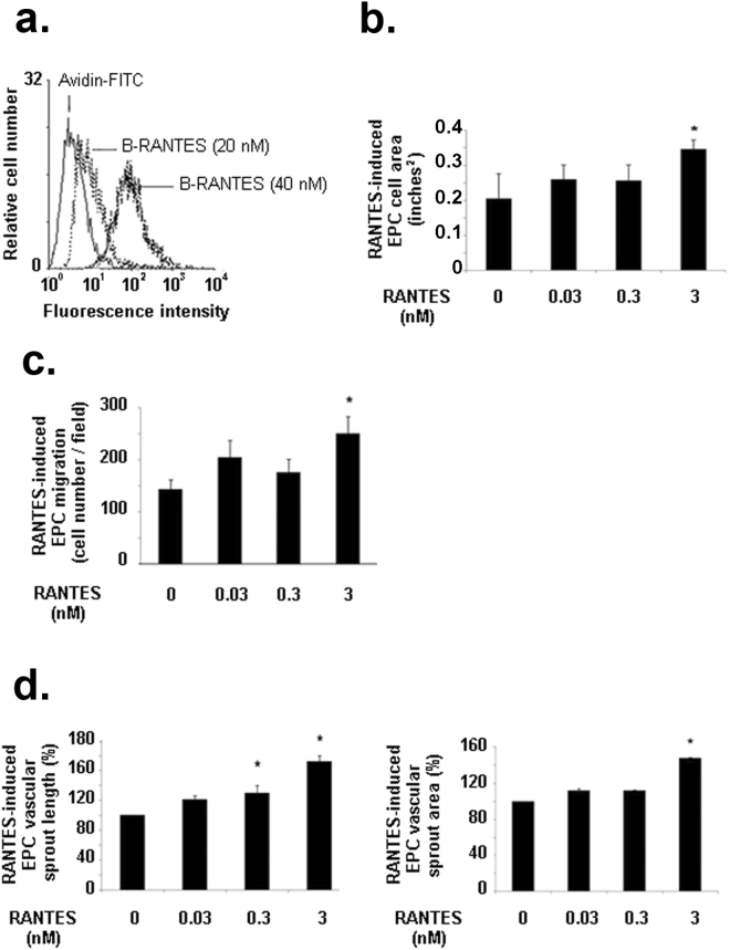
Figure 4.
Biological effects induced by RANTES on EPC. (a) RANTES binds to EPC in a dose-dependent manner. Human EPC were incubated with 20 or 40 nM biotinylated RANTES (B-RANTES) and the binding were analysed by flow cytometer with avidin-FITC. Reactivity was compared to avidin-FITC alone. Data shown are representative of three independent experiments. (b) RANTES at 3 nM induced EPC spreading on fibronectin layer. Results are expressed as mean ± SEM of EPC area, expressed in square-inches, measured by field for three independent experiments. *P < 0.05 versus untreated cells. (c) RANTES at 3 nM induced EPC migration in a modified Boyden chamber model. Results are expressed as mean ± SEM of EPC counted by field for three independent experiments. *P < 0.05 versus untreated cells. (d) RANTES at 3 nM induces EPC vascular sprout area (left panel) and vascular sprout length (right panel) and in a 2D angiogenesis assay on Matrigel. The length and area of vascular sprouts formed by untreated EPC was arbitrary set to 100%. The results for cells treated with RANTES were expressed as a percentage of control cells. *P < 0.05 versus untreated cells.

