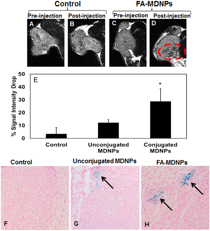
Figure 4.
In vivo MRI potential of MDNPs. (A,B,C,D) MRI images of the tumors in the control group and the group treated with folic acid-conjugated MDNPs (FA-MDNPs) before and after injection of the respective solutions. A distinct darkening of the tumor was observed in the group treated with MDNPs post injection. (E) Significant T2 signal intensity drop was observed in the case of FA-MDNPs indicating greater negative contrast due to the presence of iron oxide in the tumor (n = 4). (F,G,H) Prussian blue staining on the tumors (10x magnification). More blue regions (arrows) seen in the FA-MDNPs group indicating presence of greater amount of iron oxide in the tumor.

