Fig. 2.

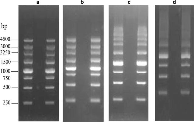
Agarose gel electrophoresis patterns of DNA. Agarose from a Biowest, b G. asiatica, c G. lemaneiformis, and d G. bailinae. The gels were exposed to UV light and the picture were taken with a gel documentation system

Agarose gel electrophoresis patterns of DNA. Agarose from a Biowest, b G. asiatica, c G. lemaneiformis, and d G. bailinae. The gels were exposed to UV light and the picture were taken with a gel documentation system