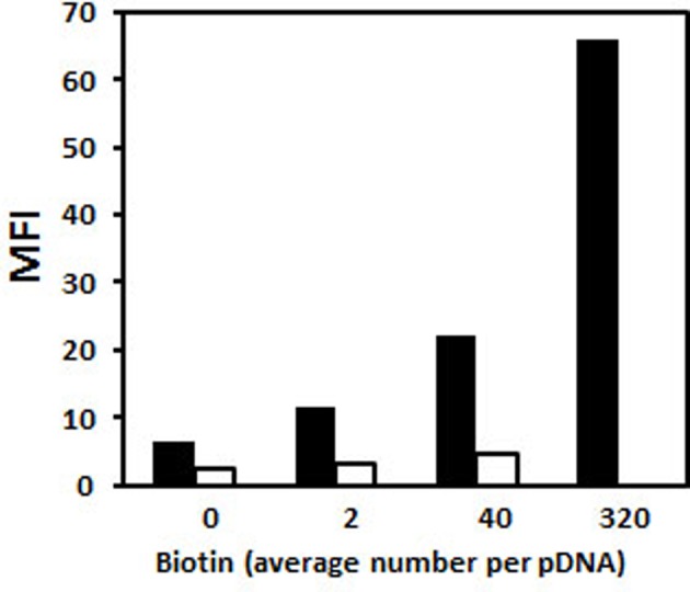
Figure 14. Biotin accessibility within polyplexes and lipoplexes.

pDNA (pCMV-eGFP) was substituted with various amounts of biotin residues. Polyplexes were formed with His–lPEI (black bar) at DNA/polymer weight ratio of 1/6. Lipoplexes were formed with KLN25/MM27 liposomes (white bar) at DNA/lipid weight ratio of 1/2. DNA complexes were mixed with fluorescein-labelled streptavidin and then the fluorescence intensity of polyplexes and lipoplexes was measured by flow cytometry.
