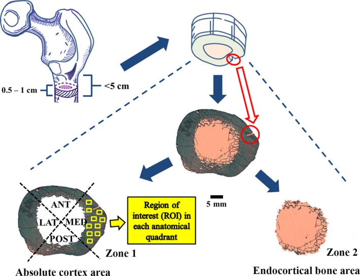
Figure 1.

Cross‐sectional femoral shaft (thickness: 5–10 mm) extracted from the subtrochanteric region (between lesser trochanter and 5 cm distally) of proximal femur were indicated with dashed lines and a minor cut was circled in red on the medial bone edge. The entire cross‐sectional femoral shaft was scanned to acquire a complete histological image (×50), which was separated into two independent zones: the absolute cortex area and the endocortical bone area. The former showed four anatomical quadrants: medial, posterior, lateral and anterior shaft. In each of them, 10 regions of interests (ROI: 1.51 mm2 for each) are indicated by solid yellow boxes.
