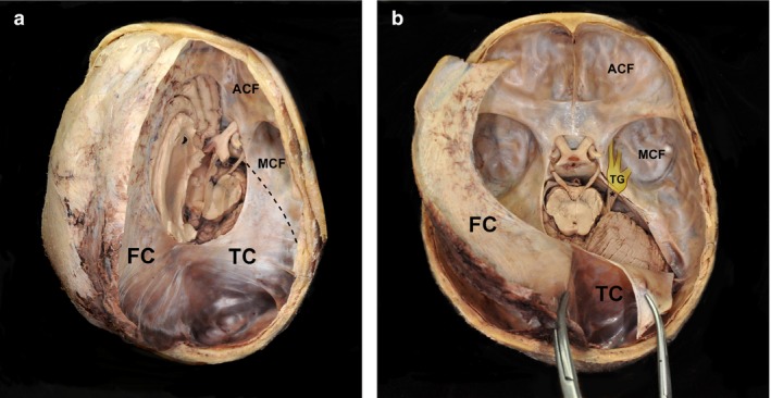
Figure 1.

Exposed cranial dura mater from a human cadaver. (a) Removing the dura mater of the right hemisphere reveals the tentorium cerebelli covering the cerebellum and attached posteriorly to the occipital bone. (b) Elevation and reflection of the anterior portion of the falx cerebri from the frontal crest and the crista galli. The lateral margin of the tentorium cerebelli based on the superior petrosal sinus is incised and reflected. ACF, anterior cranial fossa; FC, falx cerebri; MCF, middle cranial fossa; TC, tentorium cerebelli; asterisk, trigeminal nerve from the pons; dashed line, superior border of the petrous body; TG, trigeminal ganglion under the dura mater.
