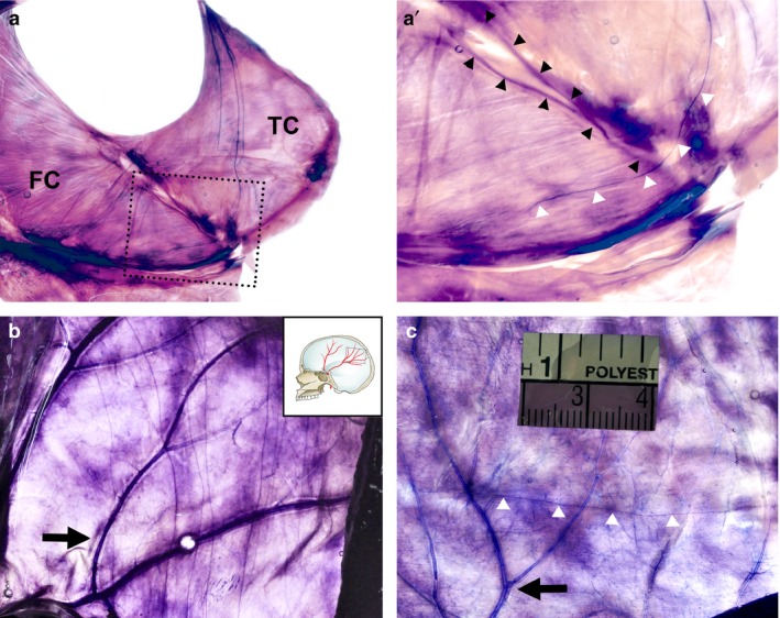
Figure 4.

Innervation of the adjacent dura mater revealed using Sihler's stain. (a) Innervation of the posterior part of the falx cerebri elongated from the tentorium cerebelli. (a') Magnification of the dotted box in (a). White triangles indicate a nerve fibre of the nervus tentorii traversing to the straight sinus. (b) The parietal branches of the middle meningeal artery (MMA) in the lateral convexity that projects via multiple nerve fibres elongated from the tentorium cerebelli. (c) A single nerve fibre (white triangles) orthogonal to the parietal branches of the MMA in another specimen. FC, falx cerebri; TC, tentorium cerebelli; black arrows, parietal branches of the middle meningeal artery; black triangles, straight sinus; white triangles, nerve fibres.
