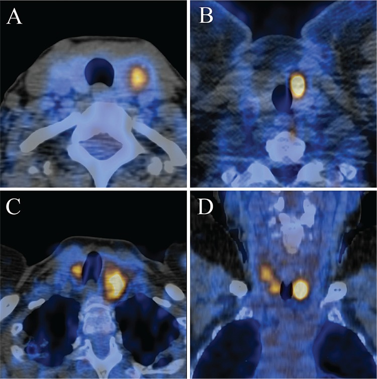
Figure 2. Four cases of incidental focal 18F-FDG uptake in the thyroid. Transaxial PET/CT fusion image of a benign left thyroid incidentaloma in a 52-year-old woman with a prior history of cervical cancer, with SUVmax 4.4, biopsied to reveal a benign follicular lesion (A), transaxial PET/CT fusion image of a malignant left thyroid incidentaloma in a 40-year-old man with a prior history of melanoma, with SUVmax 11.8, biopsied to reveal a papillary carcinoma follicular variant (B). Transaxial PET/CT fusion image of a bilateral focal benign thyroid incidentaloma in a 65-year-old woman with a prior history of lymphoma, with SUVmax 7.8 of left lesion and SUVmax 6.4 of right lesion, biopsied to reveal benign nodular hyperplasia (C), coronal PET/CT fusion image of a bilateral malignant thyroid incidentaloma in a 70-year-old woman with a prior history of colorectal carcinoma, with SUVmax 8.1 in the left lesion and SUVmax 4.5 in the right lesion, biopsied to reveal a multifocal papillary carcinoma, classical variant on a background of Hashimoto’s thyroiditis (D).

