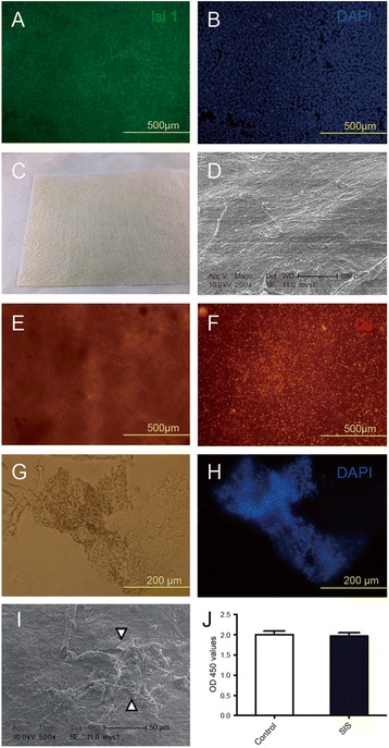
Fig. 1.

Isl1+ CPCs remain viable and attach to the extracellular matrix of the patch material (SIS-ECM). a, b Isl1+ CPCs. c Extracellular matrix (SIS-ECM) patch material. d Scanning electron micrograph of SIS-ECM. e, f Viable Isl1+ CPCs prestained by Dil in SIS-ECM obtained by bright-field (e) and fluorescence (f) microscopy. g, h Images of frozen sections of Isl1+ CPCs seeded into SIS-ECM obtained by bright-field (g) and fluorescence microscopy (h). i Scanning electron microscopy shows attachment of Isl1+ CPCs (arrowhead) in SIS-ECM. j CPC proliferation rate in SIS-ECM compared to standard culture (control) measured using the Cell Counting Kit-8 (CCK-8) assay. SIS small intestinal submucosa
