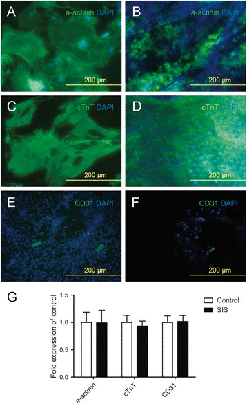
Fig. 2.

Isl1+ CPCs differentiate into CMs and endothelial cells after seeding into SIS-ECM. a, c Isl1+ CPC-derived CMs (α-actinin-positive and cTnT-positive) in culture obtained by fluorescence microscopy. b, d Isl1+ CPC-derived CMs (α-actinin-positive and cTnT-positive) in SIS-ECM obtained by fluorescence microscopy. e Isl1+ CPC-derived endothelial cells (CD31+) in culture obtained by fluorescence microscopy. f Isl1+ CPC-derived endothelial cells (CD31+) in SIS-ECM obtained by fluorescence microscopy. g Relative quantitative RNA expression of α-actinin, cTnT, and CD31 by CPC-derived CMs or endothelial cells growing in SIS-ECM compared to standard culture plates. SIS small intestinal submucosa
