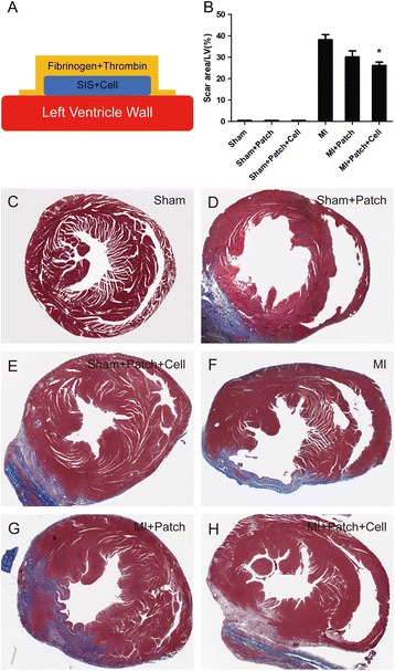
Fig. 4.

Implantation of SIS-ECM seeded with CPCs reduced myocardial scarring. Schematic view showing attachment of SIS-ECM-CPC patch to the heart with fibrin gel (a). Percent area of myocardial scarring in mice with MI. Data are mean ± SD obtained from Masson’s trichrome slides (b). Data expressed as mean ± SD. n = 6. *p = 0.003 vs MI mice. Representative images of mouse hearts (Masson’s trichrome staining) for Sham group (c), Sham + Patch group (d), Sham + Patch + Cell group (e), MI group (f), MI + Patch group (g), and MI + Patch + Cell group (h). LV left ventricular, MI myocardial infarction, SIS small intestinal submucosa
