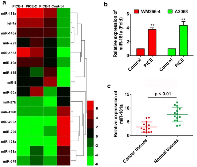
Fig. 2.

Piceatannol promotes the expression of miR-181a. a Unsupervised analysis heat map of 4 different samples. All samples are underwent cell clustering, and the top 16 microRNAs with highest standard deviation (SD > 1) are enlisted. Each column represents one specimen and each row represents one microRNA. The color scale from 6 to − 6 shown at the right bottom of color map indicates the relative expression of a miRNA in all subjected samples: green color represents expression level lower than control while red color represents an expression level higher than control, 8 up-regulated and 8 down-regulated miRNAs were shown on the map; b Relative expression of miR-181a with and without piceatannol treatment in WM266-4 and A2058 cells; c miR-18a relative expression level in cancer tissues compared with normal tissues was analyzed using qRT-PCR. *P < 0.05, **P < 0.01 compared to normal control
