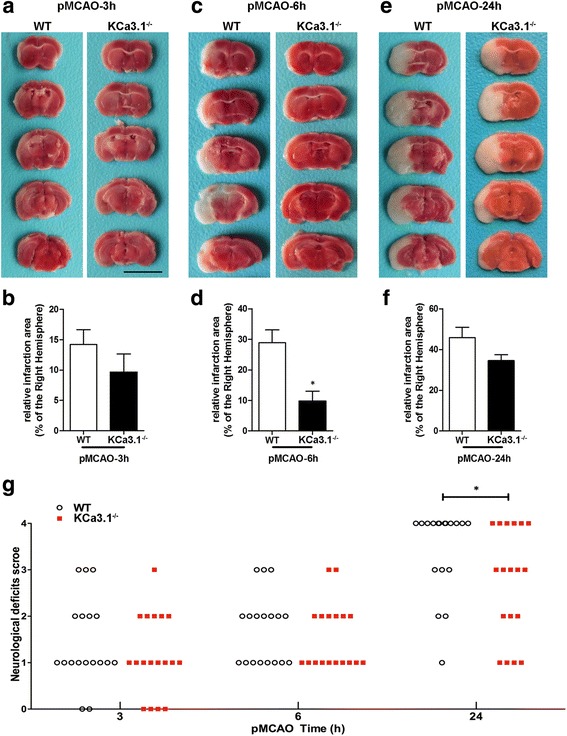
Fig. 2.

KCa3.1 deficiency reduces infarction volume and improves of neurological conditions. Focal cerebral ischemia was induced by pMCAO. a, c, and e Representative TTC staining of five corresponding coronal brain sections of a 10-week-old male WT mouse and a 10 week-old male KCa3.1−/− mouse after 3 h (a), 6 h (c), and 24 h (e) of pMCAO. b, d, and f. Quantitative analysis of infarction volume in a, c, and e, respectively. Data are presented as means ± SEM. n = 6. * p < 0.05, unpaired, two-tailed Student’s t test compared with ischemic WT group. g Neurological deficits were assessed at 3, 6, and 24 h after pMCAO. Significance: * p < 0.05 vs WT group (n = 18). Mann–Whitney test compared with WT mice. The survival rate of 24 h after pMCAO was obviously higher in the KCa3.1−/− group than that in the WT group (66.7 vs 37.5%, respectively), and significant differences was observed (p = 0.0486, chi-square test)
