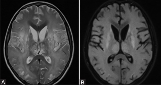
Figure 2 (A and B).

(A and B) MRI done 2 months after onset of symptoms. T2 weighted and FLAIR axial images reveal persistence of the altered signal in the deep grey matter. Also seen is presence of diffuse cerebral atrophy in the both cerebral hemispheres
