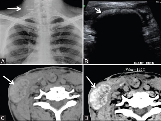
Figure 10 (A-D).

Another case of retained surgical swab in the lower neck, post neck dissection. (A) Radiograph showing irregular linear and specks of densities (white arrow). (B) Sonography image showing cavity showing highly reflective contents with posterior acoustic shadowing. (C and D) Plain and contrast-enhanced axial CT scan images show hyperdense well-defined mass showing intense but heterogeneous contrast enhancement. The central area shows negative HU due to the presence of gas. Note non-visualization of the radiodense marker
