Figure 11 (A-D).

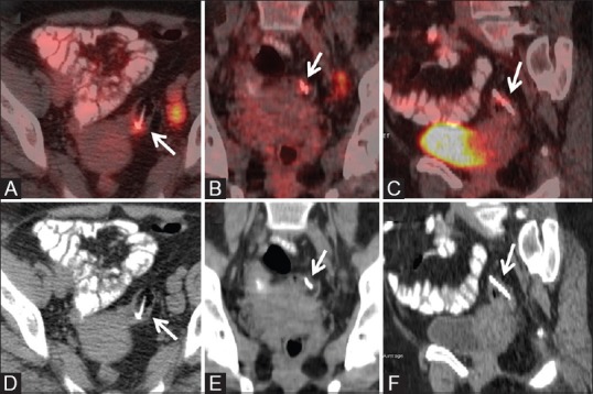
Migrated Copper T in pelvis seen on PET-CT. The axial, coronal and sagittal reformatted images of fused PETCT (A-C) and CT images (D-F) showing an FDG uptake around the foreign body which corresponds to inflammatory changes

Migrated Copper T in pelvis seen on PET-CT. The axial, coronal and sagittal reformatted images of fused PETCT (A-C) and CT images (D-F) showing an FDG uptake around the foreign body which corresponds to inflammatory changes