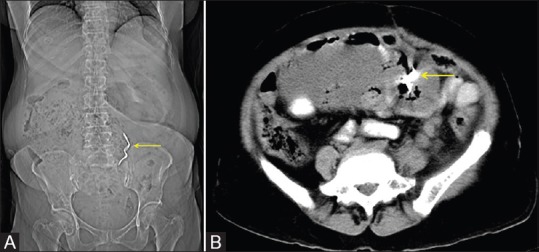
Figure 3 (A and B).

A case of carcinoma ovary, post-operative abdominal radiograph showing a linear hyperdense structure in the lower abdomen (A). The axial sections of the CT showing poorly defined fluid collection between bowel loops and a hyperdense structure within (B)
