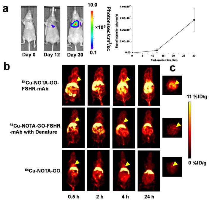
Figure 3.
Experimental murine model of breast cancer lung metastasis and in vivo PET imaging with 64Cu-labeled GO conjugates. (a) Serial bioluminescence images and BLI signal intensity from the thoracic area of mice after intravenous injection of cbgLuc-MDA-MB-231 cells; (b) Serial coronal PET imaging of cbgLuc-MDA-MB-231 tumor-bearing mice at different time points post-injection of 64Cu-NOTA-GO-FSHR-mAb, 64Cu-NOTA-GO-FSHR-mAb with denature and 64Cu-NOTA-GO; (c) The cross-sectional slices of mice containing cbgLuc-MDA-MB-231 tumor nodules at 4 h post-injection of 64Cu-NOTA-GO-FSHR-mAb, 64Cu-NOTA-GO-FSHR-mAb with denature and 64Cu-NOTA-GO.

