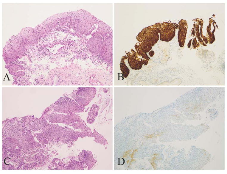
Figure 1.
Morphologic CIN 2 lesions display distinctly positive and negative p16 results. A and B, A lesion displays strong/diffuse staining (block-positive in LAST terms) and tests positive for HPV 16. C and D, A lesion displays weak, discontinuous and focal staining (negative) and tests negative for HPV (A, C: H&E, original magnification ×100; B, D: corresponding p16 IHC, original magnification ×100).

