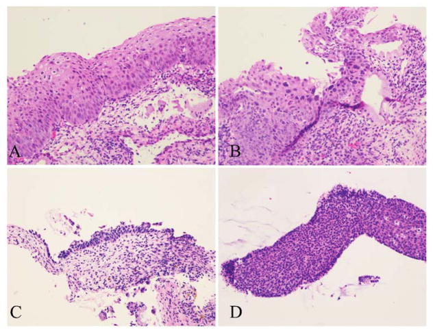
Figure 2.
Four of the most common histological features trigger the differential diagnosis of CIN 2 and deployment of p16 IHC. A, Mitoses located in the middle level and/or atypical mitoses. B, Marked koilocytic atypia. C, Eroded or thin epithelium displaying abnormal nuclear features. D, Tangentially sectioned epithelium with substantial expansion of the basal and parabasal layers (H&E, original magnification ×200).

