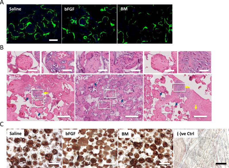
Figure 3.
Calcium deposition and histological staining for bone formation. (A) Calcein labeling suggests neo-calcium deposition in saline-, basic fibroblast growth factor (bFGF)-, and bone marrow (BM)-soaked grafts after 2 weeks of calcein uptake. Scale bar: 100 µm. (B) Hematoxylin-eosin (H&E) staining of grafts infused with saline, bFGF, and BM after 8 weeks of implantation for neo-bone tissue formation. Evidence of bone formation through endochondral ossification is evident in saline and BM groups (yellow arrows). Woven bone matrices with osteoblastic cells (blue arrows) were found in all groups. Scale bar: 200 µm. Magnified images on top are regions undergoing endochondral ossification or woven bone from regions highlighted by white borders. Scale bar: 50 µm. (C) Immunohistochemical staining for osteocalcin in saline-, bFGF-, and BM-soaked grafts after 8 weeks of implantation. Rat skin tissues were used as negative control [(−)ve Ctrl]. Scale bar: 200 µm.

