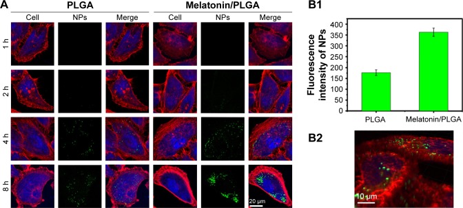
Figure 5.
(A) Cellular uptake efficiency of both PLGA and melatonin/PLGA NPs at concentration of 100 μg/mL in MCF-7 cells. PLGA was designed as the control to examine the targeting effect of melatonin on MCF-7 cell line. The images were taken by laser scanning confocal microscope (LSM780). Cytoskeleton was stained by rhodamine–phalloidin (red), the nuclei of cells were stained by DAPI (blue) and coumarin-6 was loaded in Melatonin-MNPs to track the NPs (green). (B1) The fluorescence intensity of NPs (green) after 8 h incubation with MCF-7 cells; (B2) three-dimensional images from different perspectives of MCF-7 cells using the Z-stack model of laser scanning confocal microscope.
Abbreviations: MNPs, magnetic nanocomposite particles; NPs, nanoparticles; PLGA, poly(lactic-co-glycolic acid).

