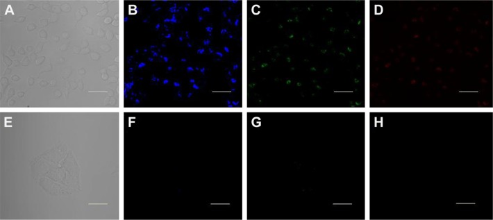
Figure 9.
The confocal fluorescence microscopy images of HeLa cells treated with 0.2 mg/mL CQDs for 4 h, (A–D) in the absence of Fe3+ and (E–H) in the presence of Fe3+ (100 μM). Images were taken under (A, E) a bright-field, (B, F) 405 nm excitation, (C, G) 458 nm excitation, and (D, H) 514 nm excitation. Scale bar =20 μm.
Abbreviations: CQDs, carbon quantum dots; HeLa, human cervical cancer cells.

