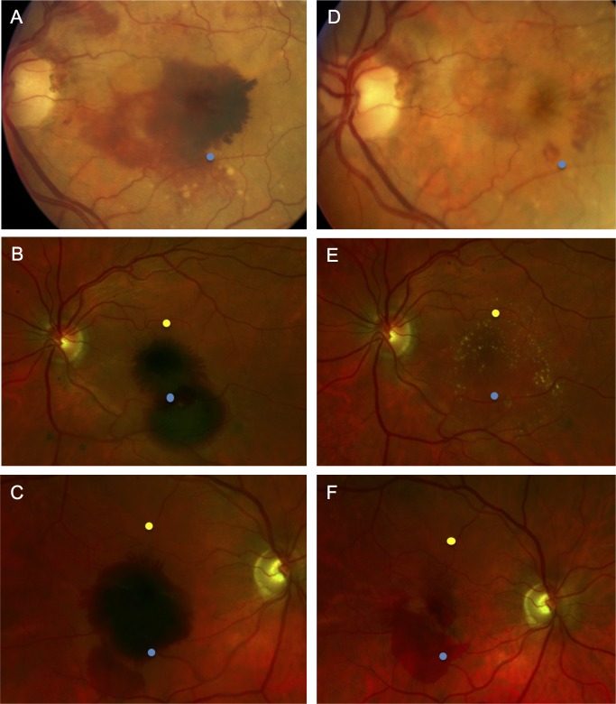
Figure 3.
Schematic of the fundus locations where OCT measurement were made before and after subretinal tPA displacement of SMH. (A–C) Fundus photographs demonstrate large SMH. (D–F) Photographs demonstrate resolution of the hemorrhages following surgery and SMH displacement with subretinal tPA at 13, 11, and 1 months, respectively. The blue points represent the location of OCT retinal thickness measurements overlying the hemorrhage, and the yellow points represent the location of measurements made immediately adjacent to the hemorrhage (attached retina). Normal, attached retinal OCT sections were not available in regions immediately adjacent to the SMH for the eyes in A and D, so yellow points are not depicted.

