. 2017 Oct 4;6:e25281. doi: 10.7554/eLife.25281

Figure 4. The anti-proliferative activity of PTX is retained following conjugation with dPG or dPGS nanocarriers.

(A) Flow cytometry analysis of cell cycle following treatment with dPG-PTX, dPGS-PTX or free PTX at different time points. Images are representative of 3 individual experiments. (B) Representative confocal images of cells treated with dPGS-PTX for 8 hr. The nucleus is stained with DAPI (blue) and tubulin is stained with FITC-labeled antibody (green). (C–E) U-87 MG and U251 human glioblastoma cells and human umbilical vein endothelial cells (HUVEC) were incubated with serial concentrations of dPG-PTX, dPGS-PTX or free PTX for 72 hr and growth inhibition was evaluated. Data represent mean ± s.d. of triplicate wells. Graphs are representative of 3 individual experiments.

Figure 4—source data 1. Raw data of U-87 MG, U251 and HUVEC proliferation assays.
DOI: 10.7554/eLife.25281.013

Figure 4.

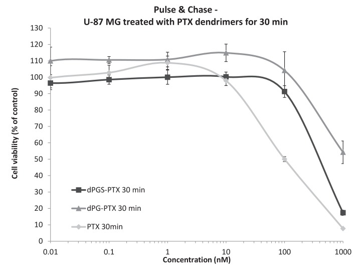
Figure 4—figure supplement 1. Differences in the in vitro activity of the dendritic conjugates are attributed to differences in their internalization kinetics.

Figure 4—figure supplement 1.

U-87 MG cells were incubated with serial concentrations of dPG-PTX, dPGS-PTX or free PTX for 30 min, washed, left to grow for additional 72 hr and analyzed for growth inhibition. Data represent mean ± s.d. of triplicate wells. Graph is representative of 3 individual experiments.
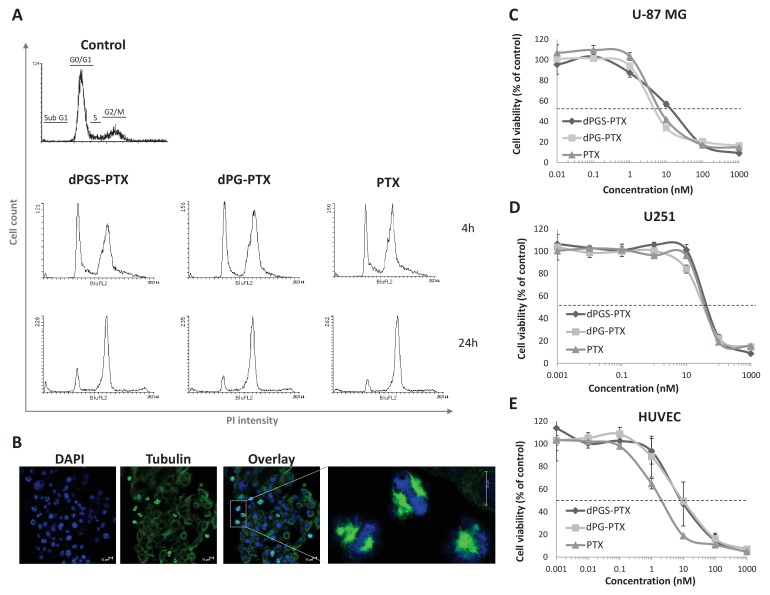
Figure 4—figure supplement 1—source data 1. Raw data of U-87 MG proliferation assay following short-term exposure to treatments.
DOI: 10.7554/eLife.25281.014