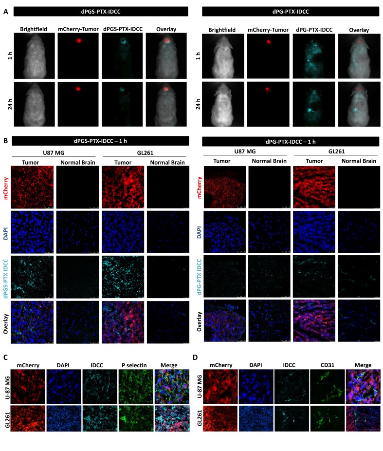
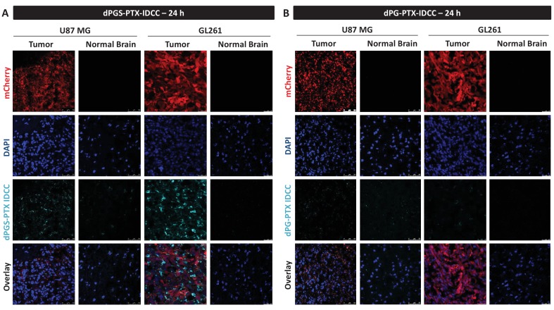
Figure 5. dPGS-PTX preferably accumulates in intracranial tumors.
(A) Representative non-invasive fluorescence images of mice bearing mCherry-labeled U-87 MG tumors (red) 1 hr and 24 hr following intravenous injection of IDCC-labeled dendritic conjugates (cyan) (n = 2). (B). Representative confocal images of brain sections of mice bearing U-87 MG or GL261 tumors 1 hr following intravenous injection of the dendritic conjugates (n = 2–3). Images depict DAPI-stained nucleus (blue), mCherry-labeled tumor cells (red) and IDCC-labeled dendritic conjugates (cyan). (C–D) Representative immunohistochemical staining for P-selectin (C) or CD31 (D) in U-87 MG and GL261 tumors following administration of dPGS-PTX-IDCC. Positive staining is shown in green (n = 3).


