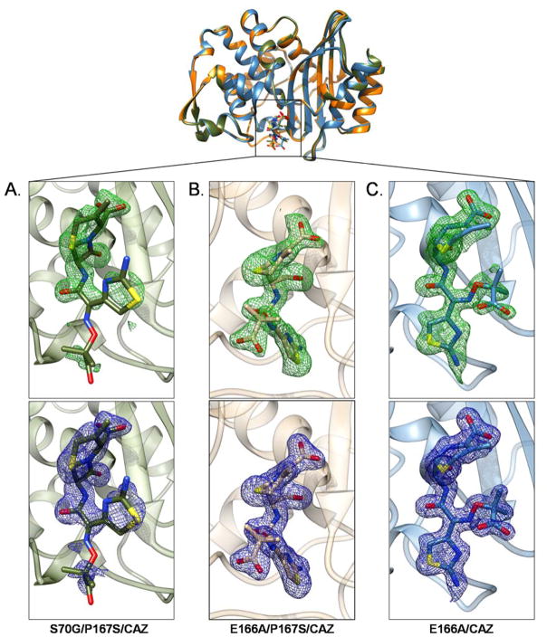
Figure 3.
Electron density for ceftazidime in the crystal structures of A) CTX-M-14 S70G/P167S/CAZ, B) E166A/P167S/CAZ and C) E166A/CAZ mutant enzymes. The alignment of the CTX-M-14 S70G/P167S/CAZ, E166A/P167S/CAZ and E166A/CAZ mutant enzymes is shown at the top. A) The intact ceftazidime molecule displayed in the active site of S70G/P167S/CAZ. Partial density is present for ceftazidime. B) The acylated ceftazidime molecule displayed in the active site of E166A/P167S/CAZ. C) The acylated ceftazidime molecule displayed in the active site of E166A/CAZ. For each of the crystal structure Fo-Fc (2.5 σ) and 2Fo-Fc (1.0 σ) maps are shown in green (top) and blue (bottom), respectively. In all panels, ceftazidime is shown as a stick model with oxygen atoms represented in red, nitrogen atoms in blue and sulfur atoms in yellow.

