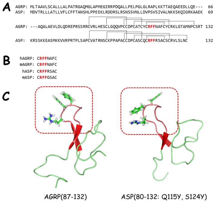
Figure 1.
A) Sequence alignment of human ASP and AGRP. The hypothesized Arg-Phe-Phe pharmacophore region is highlighted in red. The disulfide pairing is also indicated.
B) Sequence alignment of the postulated active loop of human and mouse ASP and AGRP. The conserved Arg-Phe-Phe is highlighted in red.
C) NMR solution structures of human AGRP(87-132) (PDB = 1HYK)50 and human ASP(80-132: Q115Y, S124Y) (PDB = 1Y7K).29 The β-hairpin loops are colored red and highlighted in the red box. The Arg-Phe-Phe tripeptide side-chains are drawn to illustrate their similar positions within the structures.

