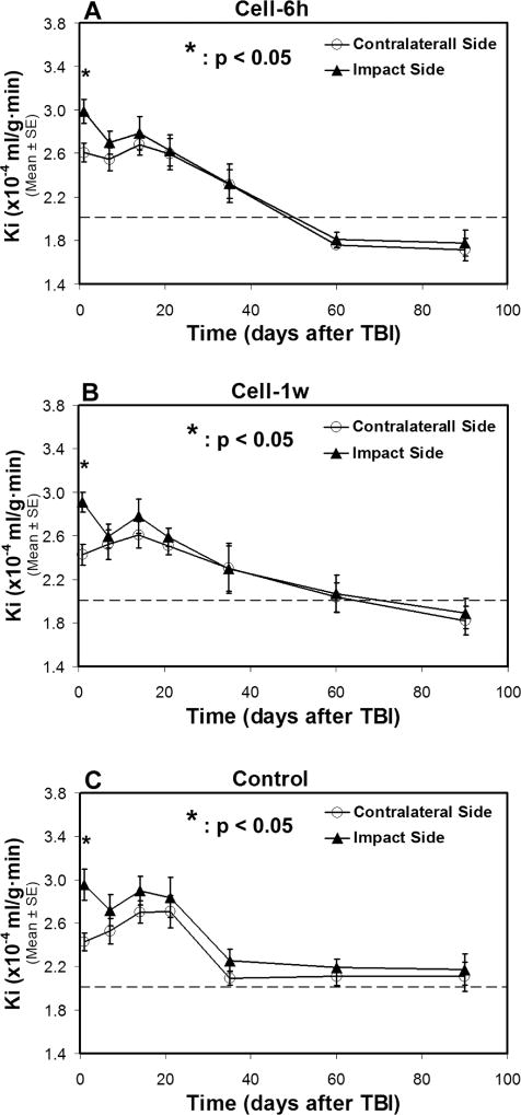
Fig. 3. Changes of Ki in two sides of normal-appearing brain tissue with (A–B) and without (C) cell administration after TBI.
A statistical difference in Ki between two sides of the brain is detected at 1-day after TBI for all treatment groups, with a significantly higher Ki value being present in the ipsilateral side than in the contralateral side of the injured brain. After 1-week post-TBI, similar temporal profiles of Ki, however, are found in two sides of the brain without statistical difference for each group, even though each group exhibits a distinct temporal pattern of Ki. The dotted line represents the Ki value in the normal rat brain.

