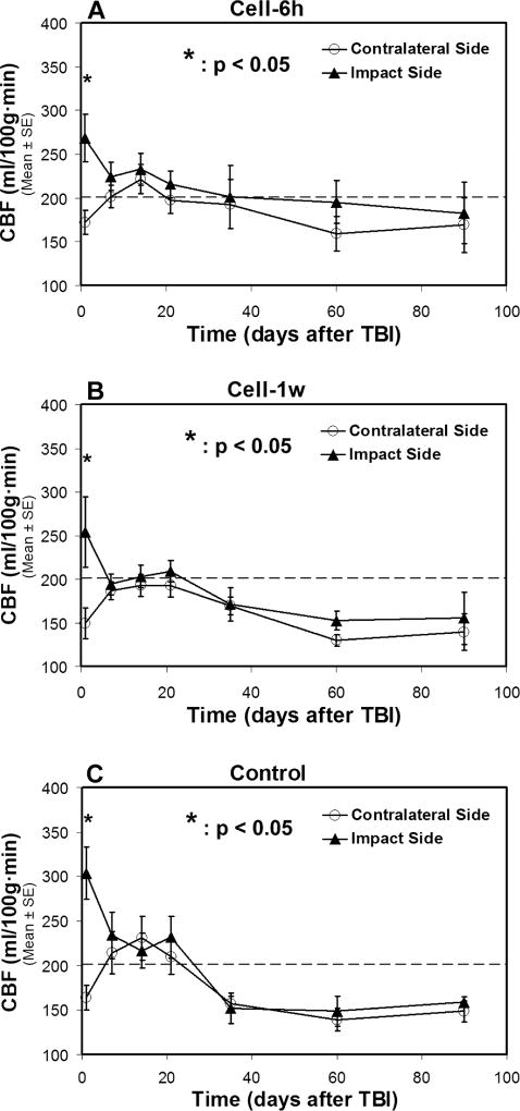
Fig. 4. Changes of CBF in two sides of normal-appearing brain tissue with (A–B) and without (C) cell administration after TBI.
A statistical difference in CBF between two sides of the brain is found at 1-day after TBI for all treatment groups, with a significantly increased CBF value being detected in the ipsilateral side than in the contralateral side of the injured brain. After 1-week post-TBI, there is no statistical difference in CBF between two sides of the brain for each group, even though the distinctive features are observed in the temporal profile of CBF for particular treatment group. The dotted line represents the CBF value in the normal rat brain.

