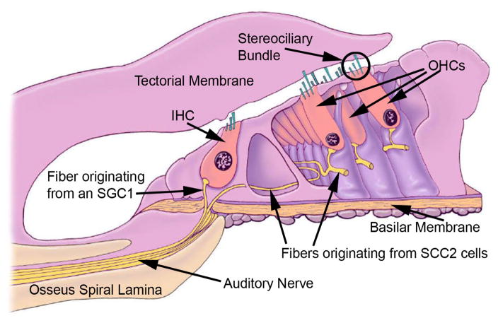
Figure 3.
A cross section of the organ of Corti showing inner hair cells (IHCs) and OHCs with their stereociliary bundles. An OHC ste-reociliary bundle is circled. The central axis of the cochlear spiral is to the left of the drawing. An IHC is located over the bony (osseus) spiral lamina and is tightly enveloped with supporting cells. Sound-evoked vibrations will be reduced at this location relative to those occurring under the OHCs located nearer the middle of the compliant basilar membrane. The mottled dark circle in the hair cells is the cell nucleus. Note the large fluid spaces around the OHCs created by the absence of adjacent supporting cells that surround the IHCs. Auditory nerve fibers (eighth cranial nerve) contact hair cells at the end opposite their mechanosensory stereociliary bundle. More than 15 fibers contact the base of each IHC while a few course laterally and each contacts more than 15 OHCs. The fibers innervating the IHCs come from type 1 spiral ganglion cells (SGC1), whereas those innervating the OHCs come from type 2 SGCs (SGC2).

