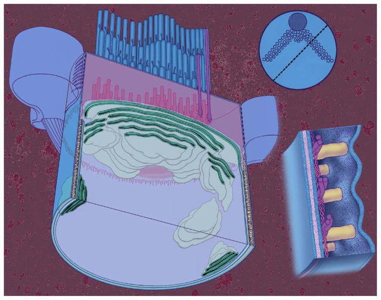
Figure 4.
The apical end of an OHC showing organization of the membranes and cytoskeletal structures. Left: a cylindrical OHC has been sliced parallel to its long axis along a plane defined by the dashed line in the insert at the top right. Top right: a view of the OHC looking down on the stereocilia bundle that has a typical W shape. Each small circle represents a single stereocilium, while the large circle represents a structure that disappears during development. Bottom right: high-power view of the lateral wall showing its three layers. The outermost layer is the plasma membrane and the innermost layer is made up of the subsurface cisterna. Sandwiched in between is the cortical lattice composed of circumferentially oriented actin filaments and axially oriented spectrin (spectrin filaments are the thinner filaments). The plasma membrane and cortical lattice form nanoscale motor elements that generate force at acoustic frequencies, resulting in electromotility.

