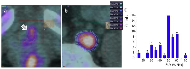
Fig. 2.
Lesion segmentation. The region of interest and segmentation of maximum standardized uptake value (SUV) with the region is shown for (a) a prostate bed lesion and (b) a lymph node lesion. (c) A histogram of threshold frequencies selected across all patients, with 50% of standardized uptake value (SUV)max being the most common value used for segmentation.

