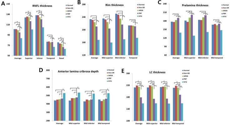
Fig 2.
A-E. Comparison of optic disc and lamina cribrosa (LC) thickness in eyes that had undergone panretinal photocoagulation (group VI) with eyes that had not (groups I, II and III) and with eyes that had normal tension glaucoma in 3–4 quadrant areas. *p < 0.05 compared with the PRP group by ANOVA post-hoc analysis using the Scheffe method.

