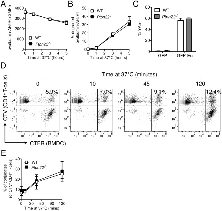
Fig 4. Ptpn22 is dispensable for antigen degradation and presentation.
(A-B) Wild type (WT) and Ptpn22-/- BMDC were incubated with ovalbumin-AF594 coated beads for 0–5 hours at 37°C. Non-internalised beads were excluded by staining with rabbit anti-ovalbumin followed by F(ab’)2 anti-rabbit-AF647. Cells were lysed and the fluorescent intensity of AF594+ AF647- beads determined by flow cytometry. (A) Ovalbumin-AF594 Geometric Mean Fluorescent Intensity (GMFI). N = 2 independent experiments ± s.d. (B) Proportion of internalised beads from WT and Ptpn22-/- BMDC that have reduced ovalbumin-AF594 fluorescence. N = 2 independent experiments ± s.d. (C) WT and Ptpn22-/- BMDC were incubated with GFP or GFP-Eα for 18 hours followed by staining for Eα52–68 in I-Ab (YAe). The percentage of YAe+ live, singlet, CD11c+ BMDC was determined by flow cytometry. N = 3 independent experiments; bars represent mean ± s.d. (D-E) CellTrace Violet (CTV) labelled WT CD4+ OT.II T-cells were incubated with CellTrace Far Red (CFTR) stained OVA323-339 peptide pulsed LPS matured WT or Ptpn22-/- BMDC for 0–120 minutes at 37°C, and the proportion of CTV+ CTFR+ conjugates within the CTV+ T-cell population determined by flow cytometry. (D) Representative flow cytometry plots showing conjugates (top, right hand quadrant, CTV+ CTFR+). (E) Proportion of DC:T-cell conjugates within CTV+ gate. N = 4 independent experiments; line represents mean ± s.d. Data (C) determined non-significant by unpaired T-test. Differences between genotypes in (E) were deemed non-significant by two-way ANOVA with Sidak’s Multiple comparison test.

