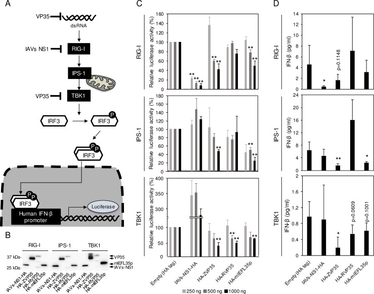
Fig 4. Inhibition of the RIG-I-mediated signaling pathway by mlEFL35 and VP35s.
(A) The RIG-I-mediated signaling pathway is shown. The human IFN-β promoter is activated through RIG-I, IPS-1 and TBK1. The IFN-β promotor activity was measured by luciferase reporter assays. (B) HEK 293 cells were transfected with each plasmid expressing HA-tagged influenza A virus NS1 (IAVs-NS1-HA), EBOV VP35 (HA-ZVP35), RESTV VP35 (HA-RVP35) or mlEFL35p (HA-mlEFL35p) and the plasmids for the reporter gene expression along with the RIG-I CARD domain vector, IPS-1 or TBK1 expression vector. NS1 and VP35 are known as IFN antagonists. Western blotting was performed to examine the expression of NS1, VP35s, and mlEFL35p. Each HA-tagged protein (IAVs-NS1-HA, HA-ZVP35, HA-RVP35, and HA-mlEFL35p) was detected with an anti-HA-tag antibody. (C) Transfected cells were solubilized and luciferase assays were performed. Relative luciferase activities were calculated by setting the values given by the cells transfected with a control empty plasmid expressing the HA tag alone. Significantly lower values compared to control cells (Empty) are indicated by asterisks (*p < 0.05, **p < 0.01). (D) Concentrations of IFN-β in the supernatants of cells transfected with the indicated plasmids (1000 ng) were measured by ELISA. Means and standard deviations of five independent experiments are shown. Significantly lower values compared to control cells (Empty) are indicated by asterisks (*p < 0.05, **p < 0.01).

