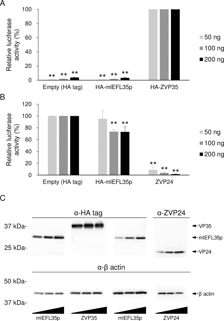
Fig 5. Luciferase expression from the Ebola virus minigenome with mlEFL35p.
(A) HEK 293T cells were transfected with the indicated amounts of plasmids for the expression of the HA tag alone, HA-tagged mlEFL35p (HA-mlEFL35p), or EBOV VP35 (HA-ZVP35) along with plasmids for the expression of NP, VP30, L, the T7 polymerase and p3E5E-luc. Relative luciferase activities were determined by setting the values of control cells transfected with the HA-ZVP35-expressing plasmid to 100%. Means and standard deviations of three independent experiments are shown. Significant differences from control cells (HA ZVP35) are indicated by asterisks (*p < 0.05). Between the empty control and mlEFL35p, there was no significant difference. (B) HEK 293T cells were transfected with the indicated amounts of plasmids for the expression of the HA tag alone, HA-tagged mlEFL35p (HA-mlEFL35p), or EBOV VP24 (ZVP24) along with plasmids for the expression of NP, VP35, VP30, L, the T7 polymerase and p3E5E-luc. ZVP24 was used as a positive control. Means and standard deviations of three independent experiments are shown. Significantly lower values compared to control cells (Empty) are indicated by asterisks (**p < 0.01). (C) Expression of each protein was detected by western blotting. HA-tagged proteins (HA-ZVP35 and HA-mlEFL35p) were detected with an anti-HA tag antibody. ZVP24 were detected with a VP24-specific mouse antiserum produced with the synthetic peptide corresponding to amino acid positions 3–15 (KATGRYNLISPKK) of EBOV VP24. β actin were detected with an anti-β actin antibody.

