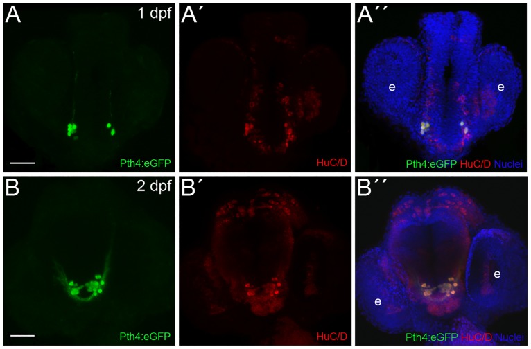
Fig 2. Zebrafish Pth4:eGFP-expressing cells are post-mitotic neurons.
Double inmunostaining in Pth4:eGFP transgenic embryos using anti-eGFP antibody (A and B) and anti-HuC/D antibody (A´and B´) shows complete co-localization at 1 and 2 dpf (A´´ and B´´). Ventral views with anterior to the top. Pth4:eGFP: green; HuC/D: red; nuclear stain: blue. Abbreviation: e, eye. Scale bar: 50 μm.

