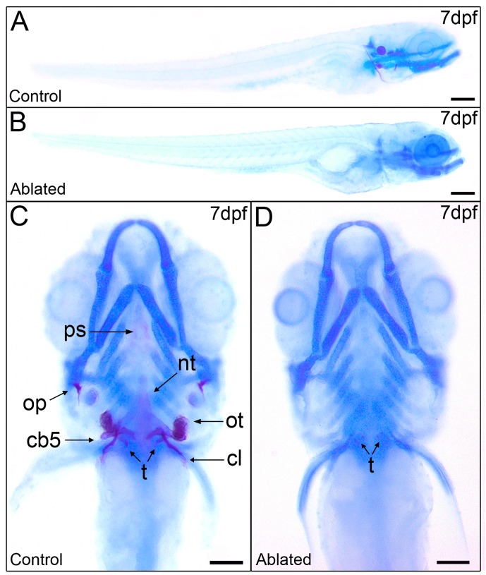
Fig 5. Ablated larvae exhibit impaired mineralization of the craniofacial skeleton but normal cartilage development.
Skeletal staining in 7 dpf control larvae (A and C) shows calcification of the craniofacial bones unlike ablated larvae (B and D) with a widely non-mineralized skeleton except an incipient mineral deposit on teeth. Lateral views (A and B) and ventral views (C and D). Abbreviations: ps, parasphenoid; nt, notochord tip; op, operculum; ot, otolith; cb5, ceratobranchial arch 5; t, teeth; cl, cleithrum. Scale bars: 100 μm.

