
Fig. 4.
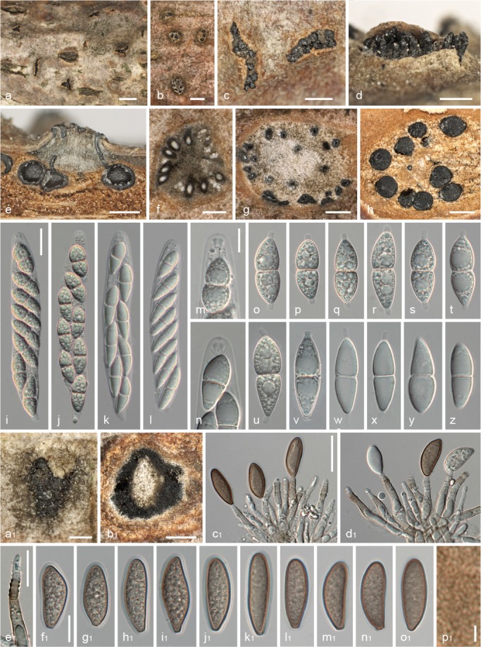
Juglanconis appendiculata. a–c. Ectostromatic discs and ostioles in surface view; d. ectostromatic disc in side view showing protruding ostioles; e. pseudostroma in vertical section; f, g. transverse sections below ectostromatic disc; h. pseudostroma in transverse section, showing perithecia and indistinct whitish to light brown entostroma; i–l. mature asci with apical ascal ring (i, j vital, k, l dead); m, n. ascus apex with apical ring (m vital, n dead); o–u. vital ascospores with cylindrical gelatinous appendages; v–z. dead ascospores (v–x showing gelatinous appendages); a1. conidioma in surface view; b1. transverse section of conidioma, showing central column; c1–e1. conidiophores (annellides) with conidia (c1, d1); f1–o1. conidia (showing gelatinous sheath in f1–k1; f1–j1 vital, k1–o1 dead); p1. detail of verruculose inner conidial wall. All in water, except k, l, n, y, z, c1–e1, l1–p1 in 3 % KOH ( a, c–e, h, u, a1, b1: WU 35955; b, g, k, l, n, w–z, c1–e1, j1–o1: WU 32010; f, i, j, o–s, f1–i1: WU 35954 (holotype); m, t: WU 35956; v: WU 35958, p1: WU 29730). — Scale bars: a = 1 mm; b, c, e, h = 0.5 mm; d, f, g, a1, b1 = 300 μm; i–l, c1–e1 = 20 μm; m–z, f1–o1 = 10 μm; p1 = 2 μm.