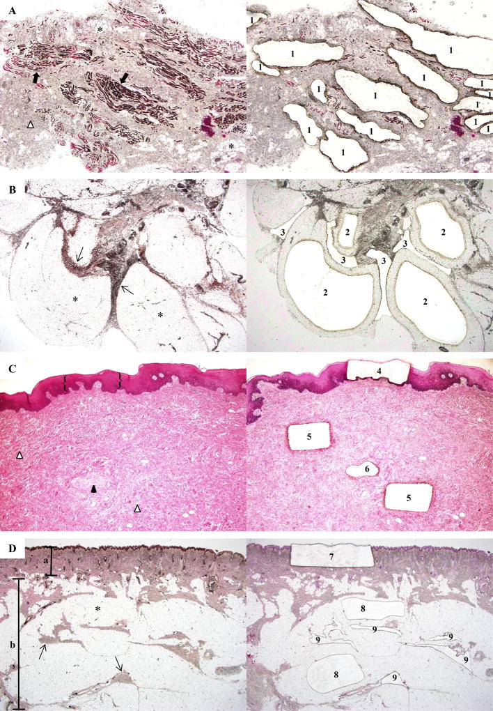
Figure 2. Laser-capture microdissection of tissue specimens from Participant 1.
(a) (Left) Orbicularis oris shows striated muscle (➡), stroma (Δ), and adipocytes (*). (Right) Post-capture image illustrates microdissection of myocyte-enriched areas (1) (microdissection of adipocytes and stromal tissue also were performed, data not shown) (x40). (b) (Left) Buccal fat lobules composed of adipocytes (*) surrounded by fibrous septa (→). (Right) Tissue section after laser-capture of adipocytes (2) and septa (3) for DNA extraction (×40). (c) (Left) Mucosal neuroma comprised of the epithelium ({}), stroma (Δ), and an enlarged nerve (▲). (Right) Tissue section after laser-capture of epithelium (4), stroma (5), and nerve (6) (x40). (d) (Left) Skin and subcutaneous tissue section show the epidermis and dermis (α), adipocytes (*) and fibrous septa (→) of the subcutaneous tissue (β). (Right) Post-capture of the epidermis and dermis (7), adipocytes (8), and stromal tissue (9) (20×). (hematoxylin & eosin staining).

