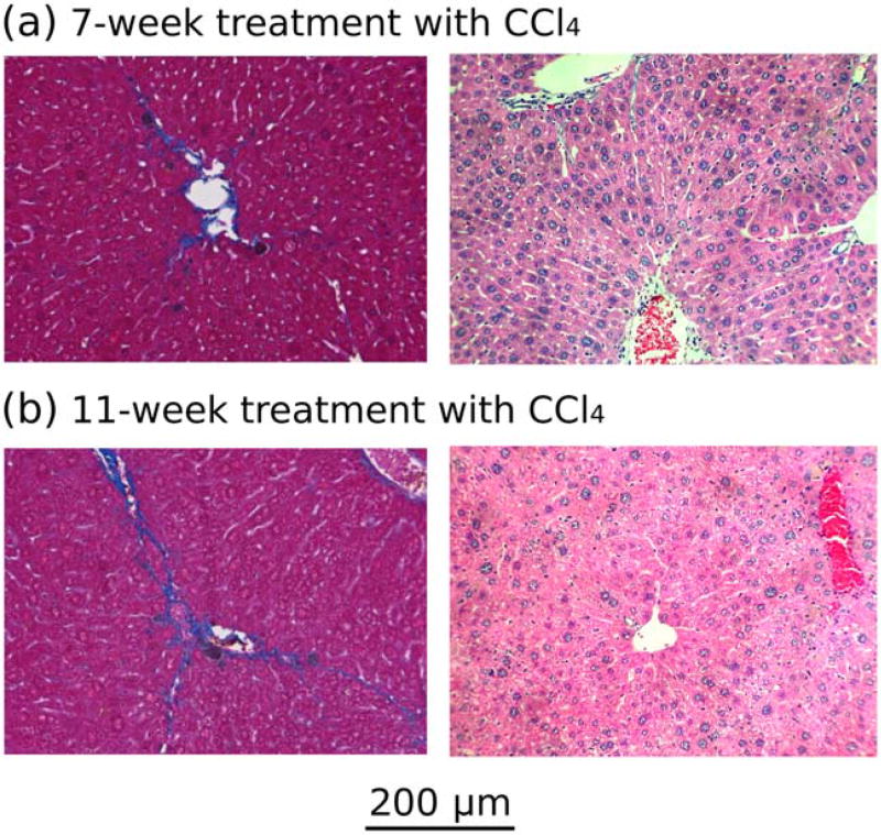
FIGURE 6.
Trichrome (left) and H&E (right) images of the livers of a representative mouse from (a) group 1 (7 weeks of CCl4 treatment) and (b) group 2 (11 weeks of CCl4 treatment). Based on a modified Brunt staging system, (a) developed fibro-sis stage 2 with centrizonal pericellular fibrosis, and (b) developed fibrosis stage 2–3 with centrizonal early septal fibrosis.

