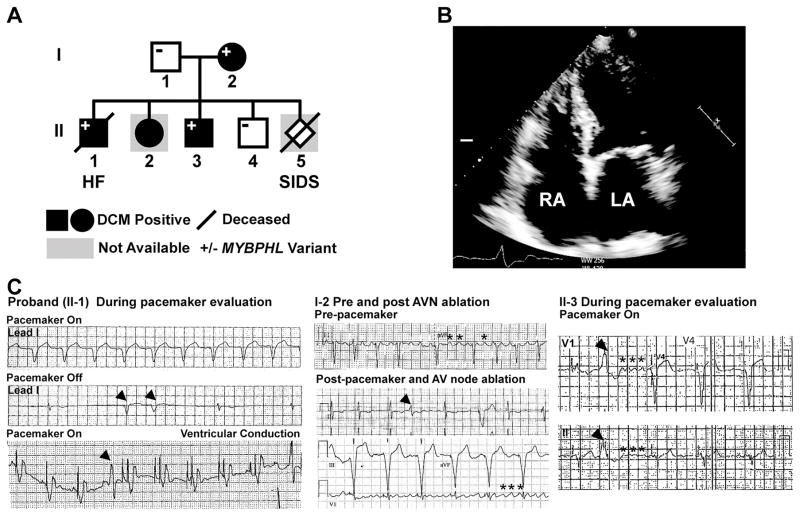
Figure 1. Whole genome sequencing (WGS) identified a premature stop codon in MYBPHL with conduction system abnormalities and DCM.
A. WGS on the proband (II-1) and parents (I-1, I-2) identified the MYBPHL R255X variant which was found in multiple family members. B. Four-chamber echocardiogram of I-2 shows enlarged right and left atria (LA, RA). C. Abnormal heart rhythms in multiple family members. ECG recordings from the proband’s pacemaker test months prior to death showing paced rhythm (top, lead 1). In the middle tracing with the pacemaker off, underlying sinus and atrioventricular node dysfunction was evident, as were PVCs (arrow heads). The bottom tracing highlights a paced rhythm and revealed PVCs (arrow head). The center panels show ECG recordings from the affected parent (I-2) show atrial flutter (asterisks) and PVC in the middle tracing. The bottom tracing shows pacing after atrioventricular (AV) node ablation with persistent atrial flutter (asterisks). The right hand panels show ECG tracing from II-3 with a PVC (arrow head) and coarse atrial fibrillation (asterisks).

