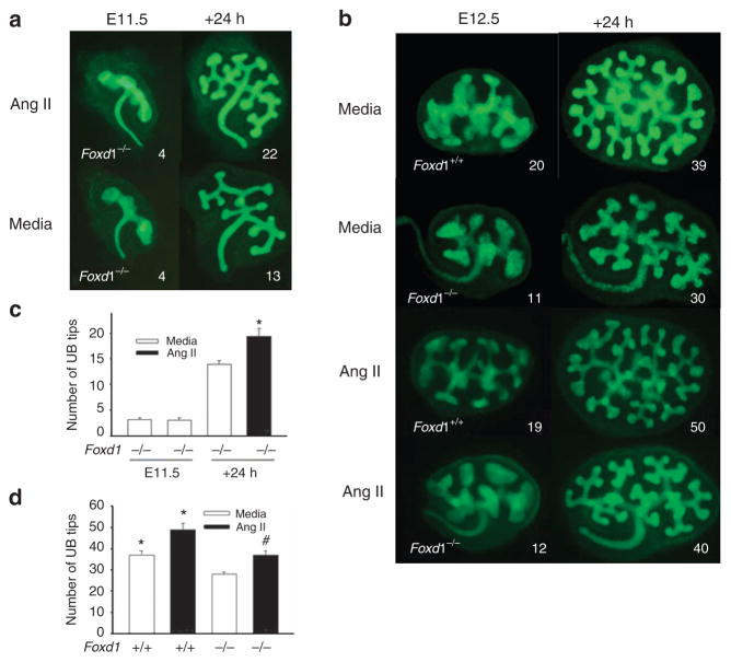
Figure 1.
Effect of genetic inactivation of Foxd1 and treatment with angiotensin (Ang) II on ureteric bud (UB) branching in mouse metanephroi. Time-lapse images of Foxd1−/−/Hoxb7GFP+ kidneys isolated on E11.5 and grown ex vivo for 24 h in the presence of Ang II (10− 5 M) or media (control; a). (b) Time-lapse images of Foxd1−/−/Hoxb7GFP+ and Foxd1+/+/Hoxb7GFP+ metanephroi isolated on E12.5 and grown ex vivo in the presence of Ang II or media (control; b). UBs are visualized with GFP (green; a,b). Bar graph showing the number of UB tips in Foxd1−/−/Hoxb7GFP+ metanephroi on E11.5 and after 24 h of culture. *P<0.05 vs. media (c). Bar graph showing the effect of Ang II or media (control) on UB tip number in E12.5 Foxd1−/−/Hoxb7GFP+ and Foxd1+/+/Hoxb7GFP+ metanephroi after 24 h of ex vivo culture (d; *P<0.01 vs. Foxd1−/−+media; #P<0.05 vs. Foxd1−/−+media). Numbers on (a,b) indicate the number of UB tips. GFP, green fluorescent protein.

