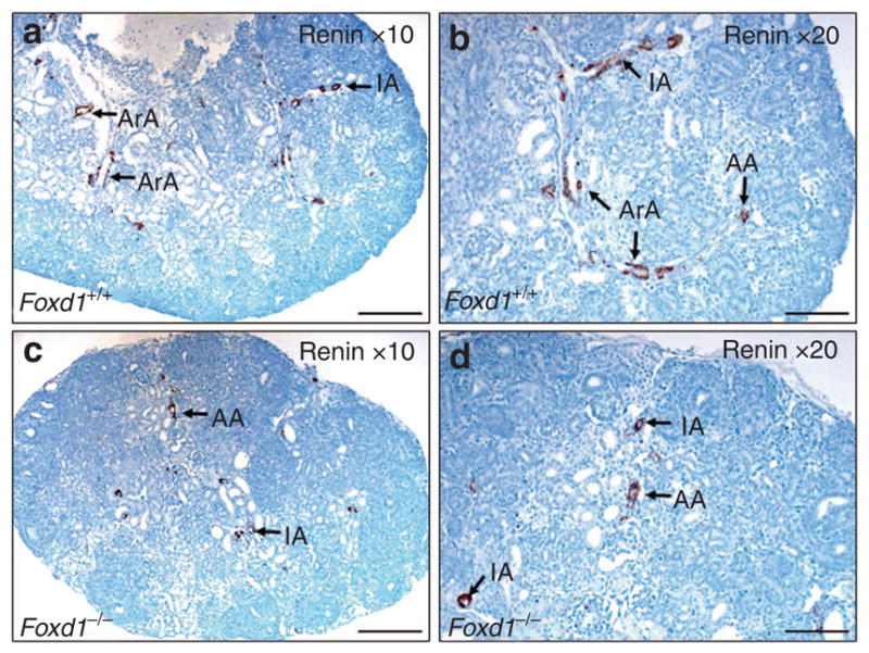
Figure 4.

Immunolocalization of renin protein in the fetal Foxd1+/+ and Foxd1−/− kidneys on E17.5. Renin immunoreactivity (brown staining) is present in the afferent arterioles (AA) and interlobular (IA) and arcuate (ArA) arteries (a,b). In Foxd1−/− kidneys, renin immunoreactivity is confined to a few cells in the vascular structures that resemble afferent arterioles and interlobular and arcuate arteries (c,d; scale bar = 100 μm).
